This article is brought to you courtesy of the National Canine Cancer Foundation.
See more articles on canine cancer.
Donate to the Champ Fund and help cure canine cancer.
Sebaceous glands are microscopic glands found below the skin. They secrete an oily substance, called sebum, which lubricates the skin and hair of animals. Tumors of the sebaceous and modified sebaceous glands are quite common in dogs. They include nodular hyperplasia, sebaceous adenoma, sebaceous ductal adenoma, sebaceous epithelioma, meibomian adenoma, meibomian ductal adenoma, meibomian epithelioma, hepatoid gland adenoma and hepatoid gland epithelioma. These tumors are usually benign in nature. But malignant sebaceous gland tumors like sebaceous carcinoma, meibomian carcinoma and hepatoid gland carcinoma too have been reported.
Dogs between 8-13 years are at an increased risk and the breeds that are genetically predisposed include English Cocker Spaniel, Cocker Spaniel, Samoyed, Siberian Husky, Cock-a-poo, Alaskan Malamute, West Highland White Terrier, Cairn Terrier, Dachshund, Miniature Poodle, Toy Poodle, Shih Tzu, Basset Hounds, Beagles and Kerry Blue Terriers. However, no sex predilection has been reported so far.
Sebaceous Adenoma, Sebaceous Ductal Adenoma and Sebaceous Epithelioma
Description
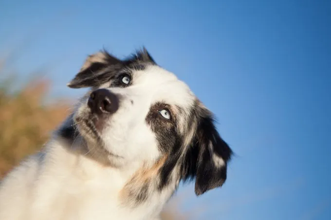
These tumors have a predilection for the head and are generally exophytic (growing outwards). But sometimes they extend into the dermis and may involve the subcutaneous tissues like the sebaceous adenomas. There are multiple lobules and at the edge of the lobules there is a reserve of small basophilic reserve cells (when a cruel disease strikes reserve cells come to the rescue of the dog) which finally differentiate into sebocytes (sebum producing cells that form the sebaceous glands).
However, sebaceous adenomas, should not be confused with sebaceous hyperplasias, which are usually multi-centric tumor like lesions found in dogs. They commonly appear as papillated (nipple like projections) masses sometimes 1 cm in diameter while sebaceous hyperplasias have a shiny, keratotic (horny growth especially like that of a wart) surface. Sebaceous adenomas are less lobulated.
Sebaceous gland epitheliomas are usually regarded a variant of sebaceous gland tumors. They are composed primarily of basal progenitor cells (they have the capacity to differentiate into a specific type of cell) rather than mature sebocytes. It is considered to be a low grade malignancy.
Sebaceous ductal adenomas consist of a large numbers of variably sized ducts which contain keratin (a tough, insoluble protein substance that is the chief structural constituent of hair, nails) and some sebum. The number of reserve cells and sebocytes seen in these tumors are few. They often have irregular lobules that extend into the deep dermis.
Cause
The exact etiology of sebaceous gland tumors is largely unknown but hormonal dysfunction could be a possible reason.
Symptoms
These tumors present themselves as elevated, nodular masses and are usually 2-5 mm in diameter. Some tumors like the sebaceous epitheliomas may appear black or brown due to the presence of melanocytes inside them. But the remaining tumors often appear as yellowish or tan on cut section and may exhibit alopecia, hyperpigmentation and ulceration with secondary infection. They may be itchy and inflamed also.
Diagnosis
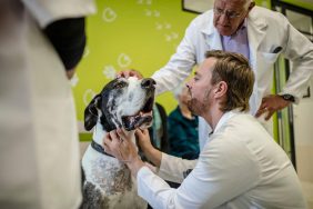
All the tumors may look alike but for accurate examination, the vet often relies on fine needle aspiration with cytology followed by histopathology and tissue biopsy.
Treatment
These tumors are amenable to surgical excision because of their benign nature. But sebaceous epitheliomas may recur twice or thrice after surgery.
Prognosis
These tumors are usually curative on surgical extirpation.
Sebaceous Carcinoma
Description
Malignant tumors of the sebaceous glands are rare in dogs and account for only 2% of all sebaceous gland neoplasms. Due to their multilobulated appearance they can be easily distinguished from a liposarcoma (malignant tumor arising in the fat cells in deep soft tissue). They are generally found on the head and neck in dogs. These tumors are locally infiltrative but rarely metastasize. Regional lymph nodes may be affected at an advanced stage of the disease.
Dogs between 9-13 years are at an increased risk and the genetically predisposed breeds may include Cocker Spaniel, West Highland White Terrier, Scottish Terrier and Siberian Husky. However, no gender predilection has been reported so far.
Symptoms – Sebaceous gland carcinomas have elevated wart like appearance and are often exophytic. They show ulceration and cause inflammation of the surrounding skin.
Diagnosis
For accurate examination, the vet often relies on fine needle aspiration with cytology followed by histopathology and tissue biopsy.
Treatment
Sebaceous gland carcinomas are amenable to wide surgical excision. However, if the tumors are found to be invasive, vets may resort to radiation therapy.
Prognosis
Sebaceous gland carcinomas usually have a guarded prognosis because of the high possibility of recurrence.
Meibomian Adenoma, Meibomian Ductal Adenoma, Meibomian Epithelioma, Meibomian Carcinoma
Description
Meibomian glands are a special kind of sebaceous glands located on the periphery of the eyelid. They are responsible for the supply of sebum which prevents the tear film from evaporating. Meibomian tumors are slow growing and may contain an excessive amount of melanin. These lesions too can be classified like the sebaceous gland tumors. However, these tumors are rare in dogs.
Meibomian carcinoma is uncommon in dogs. But they can be infiltrative and metastases via lymphatics to regional lymph nodes have been reported.
The peak incidence is between 6 and 11 and the genetically predisposed breeds include Gordon Setter, Samoyed, Standard Poodle, Shih Tzu, Siberian Husky, West Highland White Terrier and Labrador Retriever. However, so sex predilection has been reported so far.
Symptoms
Although most of them are non-malignant in nature, they generally do not metastasize. But they can cause extreme discomfort and if the tumor grows too large, it can even cause difficulties during blinking.
Diagnosis
For accurate examination, the vet often relies on fine needle aspiration with cytology followed by histopathology and tissue biopsy.
Treatment
Meibomian gland tumors are amenable to wide surgical excision. But sometimes cryosurgery (application of extreme cold to destroy deceased or abnormal tissues) may also be used. After surgery you may notice blood in your dog’s tears for a couple of days. The wound takes around 3 weeks to heal but the hair around the site will turn white permanently.
Prognosis
The tumors don’t recur often. But the first 6 months after surgery are crucial during which constant monitoring is a must.
Hepatoid Gland Adenoma and Hepatoid Gland Epithelioma
Description
Hepatoid glands are modified sebaceous glands found in skin around the anus of dogs. They are also known as circumanal glands. They derive their name from hepatocytes which are responsible for protein storage, protein synthesis and transformation of carbohydrates, synthesis of cholesterols, transformation of phospholipids, detoxification, modification and excretion of endogenous and exogenous substances. The cells of hepatoid glands resemble hepatocytes morphologically. These glands are found on the perianal region, on the dorsal and ventral aspect of the tail, in the parapreputial area in males (near the exocrine glands in front of the genitals), in the abdominal mammary region in females, on the posterior region of the hindlimbs, and on the midline of the back and thorax. Sometimes, they are found elsewhere in the body also.
Hepatoid gland adenomas are well encapsulated, multilobulated, subcutaneous masses that are benign in nature. But hepatoid gland epithelioma is poorly differentiated due to its low grade malignant nature.
Dogs between 8-13 years are predisposed and the breeds that are at an increased risk include Siberian Husky, Samoyed, Pekinese, Cock-a-poo, Cocker Spaniel, Britanny Spaniel, Lhasa Apso, Shih Tzu, Mixed Breed and Beagle. However, intact males have a higher incidence compared to their female counterparts.
Symptoms
These tumors may be found as solitary or multiple intra-dermal masses. They vary from 0.5 to 5 cm in diameter and show ulceration, alopecia and desquamation. They appear pale brown on cut section.
Diagnosis
For accurate examination, the vet often relies on fine needle aspiration with cytology followed by histopathology and tissue biopsy.
Treatment
Surgery is the treatment of choice for hepatoid gland adenomas and hepatoid epitheliomas. However, for intact male dogs, castration, (removal of the male genitals) is recommended at the time of surgery. Although chances of recurrence following surgical extirpation are minimal, fresh tumors arising from adjacent tissues are very often mistaken for recurrent hepatoid gland adenomas and epitheliomas. But hyperplastic hepatoid glands, close to the surgical margins may get transformed into new tumors at the previous site.
Hepatoid Gland Carcinoma
Description
This is a malignant tumor found on the perianal, parapreputial, and tail skin. Dogs between 8-12 years are predisposed and breeds that are at an increased risk may include Siberian Husky, Shih Tzu and Mixed Breed. Intact males have a higher predilection compared to their female counterparts.
These tumors have a variable metastatic potential and often spread via lymphatics to the sacral and iliac lymph nodes.
Diagnosis
For accurate examination, the vet often relies on fine needle aspiration with cytology followed by histopathology and tissue biopsy.
Treatment
Doctors opt for surgery followed with subsequent radiation. But hepatoid gland carcinoma is non-amenable to castration or estrogen therapy.
Prognosis
The prognosis is usually guarded in case of hepatoid gland carcinoma.
Reference
Tumors in Domestic Animals – Donald J. Meuten, DVM, PhD, is a professor of pathology in the Department of Microbiology, Pathology, and Parasitology at the College of Veterinary Medicine, North Carolina State University, Raleigh
Discover more from DogTime
Subscribe to get the latest posts sent to your email.